Latest article: enhanced second harmonic generation on nanoapertures
My team has recently published a study on second harmonic generation on single gold nanoapertures. This was released in the December 1st issue of Optics Letters. You can find a free reprint here.
Metal nanostructures are interesting emitters for second harmonic generation (SHG) radiation, which occurs essentially from the non-centrosymmetry breaking at the metal-medium interface for objects of the order of 50nm-100nm size.
At MOSAIC we demonstrate the ability of single-subwavelength-size nanoapertures fabricated in a gold metal thin film to enhance second-harmonic generation (SHG) as compared to a bare metal film. Nonlinear microscopy imaging with polarization resolution is used to quantify the SHG enhancement in circular and triangular nanoaperture shapes.
This study has two main results:
* The SHG enhancement on circular nanoapertures is demonstrated to originate from both phase retardation effects and field enhancements at the aperture edge.
* Triangular nanoapertures exhibit superior SHG enhancement compared with circular ones, as expected from their noncentrosymmetric shape.


Cool video: iso-microscopy
Isotropic single-objective microscopy (ISO-microscopy) is a novel method developed by colleagues at the Fresnel Institute to overcome the limits set by diffraction in optical microscopy. With ISO-microscopy, light is focused into an isotropic spherical spot of about one wavelength dimension.
Watch this video to learn how ISO-microscopy works !
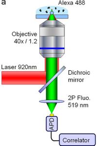
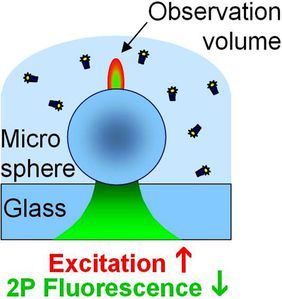
Latest article: enhanced two-photon excitation of fluorescence
Two-photon excitation of single fluorescent molecules has generated much interest and expectations, especially for analytical sciences. However, current demonstrations struggle with low two-photon fluorescence rate per molecule and/or high background. In a recent publication in Biomedical Optics Express, we answer these challenges by using a single polystyrene microsphere under focused Gaussian illumination. This opens new opportunities to extend the applications of two-photon fluorescence detection.
1- We demonstrate a simple, robust, and low-cost solution to enhance the two-photon fluorescence signal per molecule up to one order of magnitude, without adding any significant photoluminescence noise. This goes significantly beyond the current state-of-the-art.
2- We perform a thorough characterization of the two-photon fluorescence enhancement in the vicinity of a single dielectric microsphere, and report 30x higher fluorescence enhancement factors than earlier work in the field.
3- The microspheres form an interesting structure to compare the gains in one- and two-photon excitation of fluorescence. Such comparison has been seldom reported in the litterature.

In the magazine: portable single molecule fluorescence sensor
Following up with the story of the smallest single molecule detection apparatus ever, and first portable FCS system with true single molecule routine sensitivity, my team has recently published a technical note in the Spectra Analyse magazine. This is also an introduction as to why fluorescence correlation spectroscopy is a powerful method to extend standard fluorescence spetroscopies. A free reprint can be found here.
Summary: Fluorescence correlation spectroscopy (FCS) is a versatile method that would greatly benefit to remote optical fiber fluorescence sensors. However, the current state-of-the-art struggles with low detection sensitivities that prevent the extension of fiber-based FCS down to the single-molecule level. Here we describe a novel probe based on an optical fiber combined with a dielectric microsphere to perform FCS analysis down to the single fluorescent molecule level. This offers new opportunities for reducing the bulky microscope setup and extends FCS to remote or in vivo applications.
![]()

![]()
